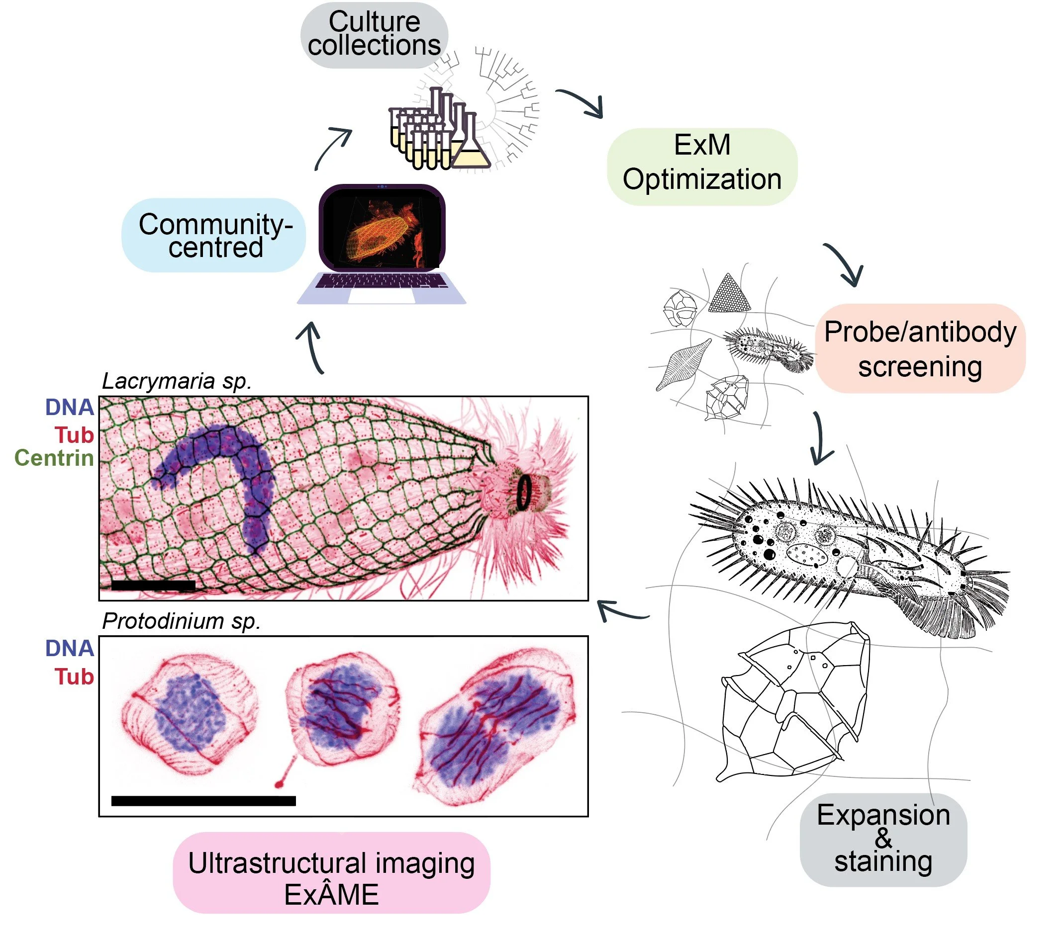
ExÂME: Expansion Microscopy Atlas of Microbial Eukayotes
Expansion Microscopy (ExM) enlarges cells by embedding them in a swellable gel, making nanoscale structures visible with standard fluorescence microscopes. In microbial eukaryotes, which are small, complex, and often shielded by tough cell walls, ExM reveals cytoskeletons, organelles, and developmental architectures in unprecedented detail, offering new insight into how these organisms grow, move, and evolve.
Supported by the Gordon and Betty Moore Foundation, ExÂME is a collaboration between the Dudin, Dey (EMBL), and Richards (Oxford) labs to build a 3D atlas of microbial eukaryotic diversity. At its heart, ExÂME is a community project that generates open imaging data, trains researchers, and makes Expansion Microscopy accessible beyond model organisms.
ExÂME carries a double meaning: it is an atlas, but also, in French, “âme”, the soul. By expanding microbial eukaryotes, we hope to glimpse something of their inner “soul”.